Did you know that you can navigate the posts by swiping left and right?
Open dataset: lumbosacral spinal cord task fMRI now on OpenNeuro
15 Sep 2026
. category:
Research
.
Comments
#neuroimaging
#fMRI
#open science
I am happy to share that the dataset underlying our Imaging Neuroscience study on lumbosacral spinal cord task fMRI is now openly available on OpenNeuro:
ds008803: Towards personalized mapping through lumbosacral spinal cord task fMRI
DOI: 10.18112/openneuro.ds008803.v1.0.0
Corresponding to: Hernandez-Charpak et al., Imaging Neuroscience, Vol. 3 (2025)
What’s in the dataset
The release covers the full acquisition from the study: anatomical, functional, timings, and physiological (cardiac, respiration) data for all 12 healthy participants across four sessions each:
- High-resolution anatomical scans: T2-weighted structural volumes of the lumbosacral spinal cord, including a reduced field-of-view ZOOMit T2w sequence for improved local resolution.
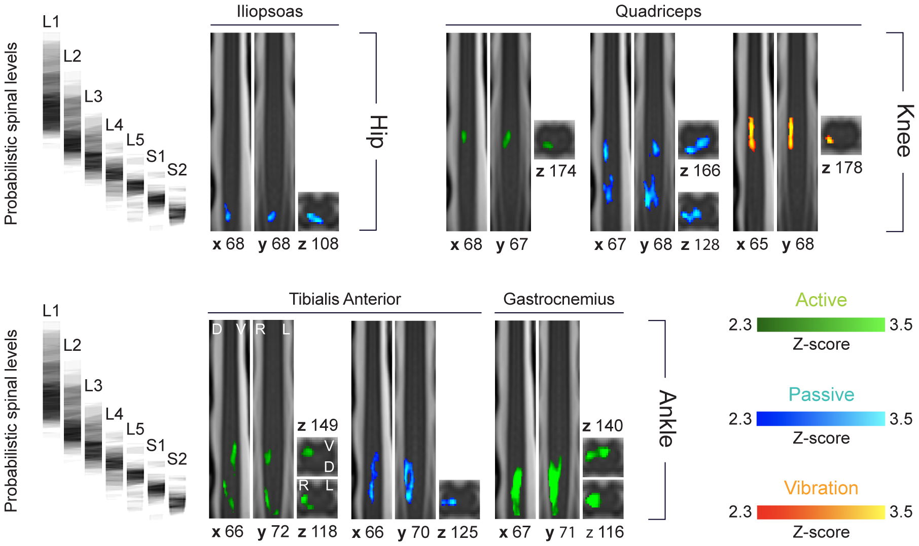
- Task-based functional MRI, spanning 14 conditions targeting the extensor and flexor muscles of the right ankle, knee, and hip:
- Ankle (4): active extension, active flexion, passive extension, passive flexion
- Knee (2): active extension, passive flexion
- Hip (2): active flexion, passive extension
- Tendon vibration (6): extension and flexion vibration at the ankle, knee, and hip, isolating proprioceptive afferent input without active or passive joint movement
- Event timing files for every run, marking stimulus/movement onsets and durations for first-level analysis.
- Physiological recordings (cardiac and respiratory traces) acquired alongside each functional run, enabling retrospective physiological noise correction (e.g. RETROICOR) — a particularly important step for spinal cord fMRI given the region’s sensitivity to cardiac-related pulsatility and respiration.
Sharing the raw data alongside the timing and physiological files means the full pipeline — from preprocessing through activation mapping — can be reproduced or re-purposed, whether to benchmark new spinal cord fMRI analysis methods, pool with other lumbosacral datasets, or revisit the active/passive/vibration contrasts with alternative models.
If you use this dataset, please cite both the OpenNeuro release and the original article linked above.
Sergio Daniel Hernandez Charpak is a PhD Computational Scientist, a Physicist and Computing Engineer researching in the interface between AI / medical imaging in healthcare, and also an enthousiast of travel, hiking, chess and piano.