Did you know that you can navigate the posts by swiping left and right?
Three abstracts at OHBM 2026 — Bordeaux
13 Jun 2026
. category:
Research
.
Comments
#neuroimaging
#fMRI
#conference
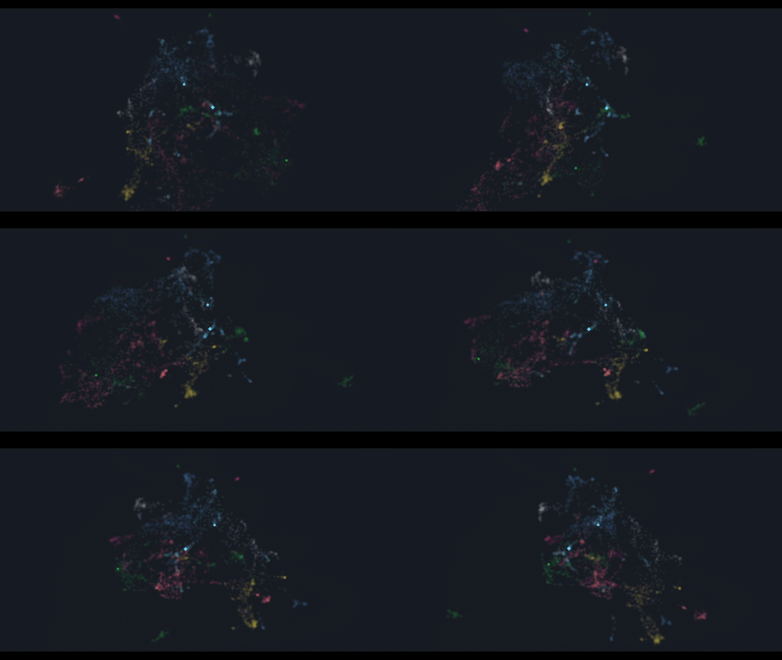
This June I attended the Organization for Human Brain Mapping (OHBM) Annual Meeting 2026 in Bordeaux, France, where I participated in three abstracts spanning spinal cord foundation modelling, resting-state functional connectivity, and proprioceptive circuit mapping. All abstracts are browsable through the Abstract Atlas — an interactive 3D embedding of every OHBM 2026 submission where each dot is an abstract, positioned by semantic similarity.
The three highlighted abstracts are visible in the visualization below: the green sphere (Abstract 0649 — SpineGPT), the blue cube (Abstract 2091 — resting-state connectivity), and the blue sphere (Abstract 0918 — proprioceptive circuits).
Abstract 0649 — SpineGPT: Self-supervised learning of human spinal cord anatomy for clinical downstream tasks
Sergio Daniel Hernandez-Charpak*, Icare Sakr*, Daniel Selmin, Léon Muller, Juliette Hars, Jonas Blanc, Bilel El-Ghallali, Philippe Forero, Jean-Baptiste Ledoux, Fabio Becce, Jocelyne Bloch#, Grégoire Courtine#, Henri Lorach#
* Contributed equally · # Contributed equally
This abstract presents the continuation of the SpineGPT framework first presented at ESMRMB 2025. Personalized spinal cord models are essential for EES therapies in SCI, Parkinson’s Disease, and MSA — but anatomical tissue segmentation from medical imaging remains a labor-intensive bottleneck for clinical scalability. SpineGPT addresses this through self-supervised learning (SSL): inspired by NLP foundation models, it learns to reconstruct randomly masked (80%) 3D chunks of T2-weighted MRI volumes, capturing the spinal cord’s underlying structure and variability without requiring labeled data.
Pre-training. The updated SpineGPT encoder (12 Vision Transformer layers, 16 attention heads, 64M parameters total) was pretrained on over 1,800 diverse T2-weighted MRI volumes spanning cervical, thoracic, lumbar, and sacral regions, drawn from both public and private datasets — up from 1,300 volumes at ESMRMB. A latent space exploration framework using T-SNE was developed to visualize and analyse the learned representations, confirming that the encoder captures region- and tissue-specific organization.
Spinal tissue segmentation. We introduced a two-stage fine-tuning strategy: first, we artificially generated a large labeled dataset (954 volumes) using a pretrained nnU-Net model for CSF, WM, and spinal roots; then we continued fine-tuning on a manually labeled high-quality dataset of 18 lumbosacral T2-weighted MRI volumes containing both ventral and dorsal spinal roots. Testing on an internal set (3 individuals) yielded Dice scores of 0.87 ± 0.21 (CSF), 0.87 ± 0.31 (WM), and 0.615 ± 0.11 (spinal roots). On a public lumbosacral benchmark (14 participants, dorsal roots only), we achieved 0.91 ± 0.03 (CSF), 0.89 ± 0.23 (WM), and 0.41 ± 0.17 (spinal roots) — matching state-of-the-art performance.
Artefact reconstruction. A new downstream task introduced at this conference: SpineGPT’s decoder is fine-tuned to identify and reconstruct MRI regions corrupted by magnetic field inhomogeneity artefacts (bending artefacts, dark-line structures). Since no ground-truth clean/corrupted pairs exist, we built a synthetic dataset by segmenting real artefacts with an nnU-Net (Dice 0.89) and generating a library of over 500 artefact shapes. Our preliminary pipeline achieved a Mean Absolute Error (MAE) of 2.251, with further improvements anticipated through adversarial training and percentage-based artefact masking.
Abstract 2091 — Potential of Personalized Spinal Cord Dynamic Functional Connectivity Across Regions and Pathologies
Olivia Ruggaber, Sergio Daniel Hernandez-Charpak, Ilaria Ricchi, Jean-Baptiste Ledoux, Fabio Becce, Jocelyne Bloch, Eduardo Martin Moraud, Grégoire Courtine, Henri Lorach, Dimitri Van De Ville
I designed this study, supervised Olivia throughout the project, and contributed to the development of the methods, the analysis and interpretation of the results. This work applies the SPiCiCAP (Spinal Cord Innovation-driven Co-Activation Patterns) framework to resting-state spinal cord fMRI in clinical populations, investigating whether intrinsic spinal activity — and its functional segmental organization — persists independently of brain input in individuals with spinal cord injury (SCI) and Parkinson’s Disease (PD). Ilaria co-supervised Olivia as well, bringing her lumbar spinal cord SPiCiCAP expertise.
Background. A key challenge for EES is determining optimal implantation sites given substantial inter-individual anatomical variability in spinal level localization. Resting-state fMRI captures spontaneous BOLD fluctuations without requiring active task performance — making it well-suited for clinical populations. SPiCiCAPs model transient co-activations through total activation deconvolution and K-means clustering, and have previously been shown to align with spinal level organization in healthy participants at cervical and lumbosacral levels.
Participants and acquisition. We studied 7 individuals with SCI (complete and incomplete, across lumbosacral, cervical, and thoracic regions) and 1 participant with PD, recruited across multiple clinical trials. Resting-state fMRI (15 min, 360 volumes) was acquired on a 3T Siemens PrismaFit with selective field-of-view ZOOMit sequences. Preprocessing included slice-timing correction, slice-wise motion correction, and physiological denoising; images were registered to the PAM50 template using subject-specific structural landmarks.
Results. To our knowledge, this is the first characterization of spontaneous spinal cord activity in SCI populations across different spinal regions. In participants with complete thoracic SCI, SPiCiCAPs demonstrated high stability (>0.8) and rostrocaudally structured activity closely aligned with spinal level anatomy — occurring entirely below the lesion, independently of brain input. This provides compelling evidence that local spinal circuits, potentially involving sensory afferent loops and interneurons, maintain organized spontaneous activity after complete supraspinal disconnection. Across incomplete SCI (cervical, thoracic, lumbosacral) and PD, SPiCiCAPs consistently revealed level-specific co-activation patterns with high cluster stability. The optimal number of clusters varied between individuals, underlining the need for personalized — rather than atlas-based — functional spinal level identification.
Abstract 0918 — Spinal functional MRI to Map Proprioceptive Circuits in Cervical and Lumbar Regions
Raphaëlle Schlienger, Caroline Landelle, Daniela Pinzon-Corredor, Sergio Hernandez-Charpak, Laurent Théfenne, Bruno Nazarian, Julien Sein, Grégoire Courtine, Jean-Luc Anton, Anne Kavounoudias
This work, led by Prof. Anne Kavounoudias, builds directly on our earlier lumbosacral and cervical spinal cord fMRI studies. By combining spinal fMRI with mechanical muscle tendon vibration — which selectively activates proprioceptive Ia afferents and induces kinesthetic illusions without actual movement — the study maps proprioceptive circuits across both cervical and lumbar spinal levels in healthy adults.
Participants and stimulation. Two independent cohorts participated: cervical (n=24) and lumbar (n=29) studies. MR-compatible pneumatic vibrators at 50–75 Hz were applied bilaterally to six muscle sites per participant (wrist flexors, biceps, anterior deltoids for cervical; anterior tibialis, quadriceps, hip flexors for lumbar). Stimulation onset was synchronized to MR acquisition via a custom LabVIEW system. Acquisitions were performed on a 3T Siemens MAGNETOM Prisma with gradient-echo EPI ZOOMit sequences (1×1mm² in-plane, 4mm slice thickness).
Results. All participants reliably reported movement illusions corresponding to muscle elongation, confirming effective proprioceptive recruitment. Group-level analyses revealed clear rostrocaudal somatotopy: cervical cord activations localized to C5 (anterior deltoid), C7 (biceps), and C8 (wrist flexors); lumbar activations peaked at L1, L4–L5 (knees and ankles), with hip stimulation engaging more rostral segments. Activations were predominantly ipsilateral and ventral, consistent with the anatomy of primary afferent projections into proprio-motor loops, though dorsal and contralateral involvement was also observed. Substantial inter-individual variability was documented — echoing clinical evidence that muscle innervation overlaps and varies between individuals. At the cervical level, this was addressed through a deep-learning-based rootlet identification method enabling individualized spinal level mapping and a population-specific probabilistic template. In the lumbar cord, ROI-based analyses were used instead.
Conclusion. Together, the two studies demonstrate that spinal fMRI combined with controlled proprioceptive stimulation can noninvasively map the organization of human spinal sensorimotor circuits across the full cervicolumbar axis, opening avenues for studying spinal plasticity and developing clinically relevant biomarkers of sensorimotor function.

Sergio Daniel Hernandez Charpak is a PhD Computational Scientist, a Physicist and Computing Engineer researching in the interface between AI / medical imaging in healthcare, and also an enthousiast of travel, hiking, chess and piano.